See also
Periorbital and orbital cellulitis
Sepsis
Antibiotics
Key points
- The typical presenting features of cellulitis include soft tissue induration, tenderness, redness, warmth and/or swelling. Large local allergic reactions are frequently misdiagnosed as cellulitis
- Most cases are mild to moderate in severity and respond well to oral antibiotics
- Marked systemic symptoms, or severe pain out of keeping with apparent severity of infection, require urgent assessment
- Oral first-generation cephalosporins are usually the first line treatment for cellulitis and other bacterial skin infections. Other factors to consider include organism exposure, MRSA risk, clinical severity, immunisation status and immunocompromise
Background
Cellulitis
- Acute bacterial infection localised to the deep dermis and subcutaneous tissue
- Predisposing factors include breaks in the skin barrier, such as abrasions, lacerations, burns, insect bites, eczematous skin, chickenpox, etc, although the portal of entry of organisms is sometimes not apparent
- The most common causes are group A streptococcus (GAS) and Staphylococcus aureus (S. aureus)
- Infections in the setting of freshwater or seawater exposure may be caused by
- Aeromonas species (fresh or brackish water, and mud)
- Mycobacterium marinum (fish tanks)
- Vibrio species (salt or brackish water, oyster shells, coral cuts)
Other common bacterial skin infections
Impetigo (commonly called "school sores")
- Highly contagious, superficial infection of the epidermis, particularly common in young children and characterised by crusty scabs. Causative organisms are GAS and S. aureus
- May also present as expanding bullous lesions
- May be associated with scabies
- Skin infections, including impetigo and scabies, are endemic in some communities in Australia, and may be associated with rheumatic heart disease and glomerulonephritis
Folliculitis/furunculosis
- Superficial (folliculitis) or deep (furunculosis or ‘boil’) infections of a hair follicle, typically presenting as one or multiple red spots or pustules
- May develop surrounding cellulitis, and can lead to abscess formation
- Often self-resolving with skin hygiene routines
- Pseudomonas aeruginosa folliculitis (‘hot tub’ folliculitis) should be considered if clusters of lesions appear following exposure to spa pool or hot tub
Infected animal/human bites
Severe presentations of bacterial skin infections
Staphylococcal scalded skin syndrome (SSSS)
- Blistering skin disorder induced by the exfoliative (epidermolytic) toxins of S. aureus
- Primarily affects neonates and young children
- Primary site of infection can be area of cellulitis, impetigo, skin trauma (burns)
- Lesions resemble hot liquid scalding burns
Necrotising soft tissue infections (NSTI)
- Rapidly progressive, characterised by necrosis of subcutaneous tissue
- High morbidity and mortality
- Causative organisms include GAS, S. aureus, and much less commonly anaerobes; may be polymicrobial
- Recent infection with varicella-zoster virus is a risk factor
There are many other forms of skin infection that are not covered in this guideline
Assessment
History
- Systemic symptoms
- Underlying skin disorder (eczema)
- Recent break in skin barrier (abrasion, burn, insect bite, animal/human bite)
- Immunisation status (Hib, tetanus)
- Overseas travel
- Water exposure (fresh, salt, mud, fish tanks)
- MRSA risk factors / colonisation
- Residence in an area with high prevalence of MRSA, eg Northern Territory, remote communities in northern Queensland
- Previous colonisation or infection with MRSA (particularly recent)
- Aboriginal and Torres Strait Islander, Pacific Islander and Māori children experience a disproportionate burden of skin infection, including MRSA-related infections. This reflects structural and social determinants of health, rather than ethnic status itself conferring risk
- Other comorbidities, eg diabetes, obesity, intravenous drug use, immunosuppression
Examination
Typical presentation of all skin infections
- Soft tissue redness (usually poorly demarcated)
- Warmth and swelling
- Pain/tenderness
Mild to moderate cellulitis
- Small to moderate area of erythema, tenderness and induration
- May present with mild systemic features, eg low grade fever in a well looking child
Severe cellulitis
- Marked swelling and/or tenderness
- Larger body surface area involved
- Tracking - painful lymphangitis (rapidly progressive, tender, erythematous streaks extending proximally up an extremity, with or without lymphadenitis)
- Palpable subcutaneous mass or induration, with or without purulent discharge, may suggest abscess formation
- Can be associated with marked systemic features, eg fever or hypothermia, tachycardia, tachypnoea, altered conscious state, unwell appearance, hypotension - late signs. See Sepsis
Features suggestive of NSTI include
- Severe pain out of keeping with apparent severity of infection
- Rapid progression with or without palpable crepitus
- Marked systemic features, eg high fever with rigors, tachycardia, tachypnoea, hypotension, confusion, vomiting. See Sepsis
Red flags
- Signs of Sepsis
- Abscess or suppuration requiring incision and drainage
- Deep structure involvement
- Immunosuppression
- Facial cellulitis in non-Hib immunised
- Foreign body
- Lymphangitis
- Secondarily infected varicella lesions
Differential diagnosis
Large local reactions such as local vaccine reactions and insect bites are a common mimic of cellulitis. Features include
- a punctum at the site
- rapid onset after bite or vaccine
- itch as a prominent feature
- redness and induration, but rarely pain
Deep vein thrombosis (DVT) of the lower limb
- Consider DVT or venous stasis in children with risk factors such as reduced mobility, increased clotting tendency, vascular abnormalities, heart failure or cancer
Management
Investigations
- Routine investigations are not indicated for mild/moderate cellulitis
- Swab for Gram stain and culture if discharge present
- Consider nasal swab if risk factors for MRSA are present or poor response to treatment
- Inflammatory markers and blood culture if severe cellulitis or suspected sepsis (blood culture is not indicated in mild/moderate cellulitis)
- Consider imaging (eg ultrasound) if abscess, deep infection or foreign body suspected
Treatment
- Manage sepsis if features present
- Manage source if identifiable (clean and debride wound, remove foreign body, drain abscess)
- Using a tool such as Melbourne ASSET score may be helpful to guide stratification of cellulitis severity and consideration of oral versus IV antibiotics
- For ongoing management refer to flowchart below
Flowchart summarising the Melbourne ASSET score and cellulitis management pathway
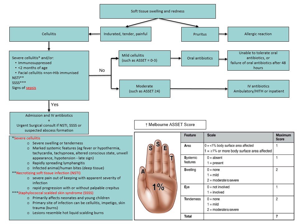
† Reproduced with permission from Ibrahim LF et al. Development and Validation of a Cellulitis Risk Score: The Melbourne ASSET Score. Pediatrics. 2019;143(2):e20181420. https://doi.org/10.1542/peds.2018-1420
Summary of antibiotic therapy
Antimicrobial recommendations may vary according to local antimicrobial susceptibility patterns. Refer to local guidelines
- Clinical features of cellulitis, in particular the associated swelling and erythema, do not usually improve significantly within 24 hours of treatment. Consider waiting 48 hours to change management
- For children with risk factors for MRSA (see above), see local antimicrobial guidelines
| Diagnosis |
Antibiotic |
Total duration |
Comments |
| Impetigo |
Topical mupirocin 2% ointment to crusted areas tds
or
Cefalexin 20 mg/kg (max 750 mg) oral tds |
5 days |
Topical mupirocin appropriate for non-recurrent, limited, non-bullous impetigo (up to 2 sores) and not at risk of acute rheumatic fever* |
| Mild cellulitis (eg ASSET score 0-3) |
Cefalexin 20 mg/kg (max 750 mg) oral tds |
5-10 days |
If oral antibiotics not tolerated or no improvement after 48 hours, manage as per moderate/severe cellulitis |
| Moderate cellulitis (eg ASSET score ≥4) or severe cellulitis |
Cefazolin 50 mg/kg (max 2 g) IV 8 hourly
orCeftriaxone 50 mg/kg (max 2 g) IV daily (for hospital-in-the-home if available)
If rapidly progressive or suspected SSSS, consider adding IV clindamycin 10 mg/kg (max 600 mg) IV 6 hourly
|
5-10 days |
When improving, switch to oral antibiotics as per mild cellulitis |
| NSTI |
Meropenem 20 mg/kg (max 1 g) IV 8 hourly and
Vancomycin 15 mg/kg (max 1 g) IV 6 hourly and
Clindamycin 15 mg/kg (max 600 mg) IV 8 hourly |
|
Urgent referral to surgical team for debridement
Seek specialist infectious disease advice for antibiotics
Consider IVIg |
| Facial cellulitis in child <5 years of age and non-Hib immunised |
Ceftriaxone IV 50 mg/kg (max 2 g) IV 12 hourly |
|
Seek specialist infectious disease advice if confirmed Hib infection |
| Mammalian/human infected bites |
See Animal and human bites |
|
|
| Waterborne skin infections |
Cefalexin 20 mg/kg (max 750 g) oral tds
and
Trimethoprim/sulfamethoxazole 8/40 mg/kg (max 320/1600 mg) oral bd
or
ciprofloxacin 10 mg/kg (max 500 mg) oral bd |
5-10 days |
Clean and debride wound as needed
|
*For children who are at risk of RHD or who live in RHD endemic areas, refer to the Australian guideline for the prevention, diagnosis and management of acute rheumatic fever and rheumatic heart disease
Consider consultation with local paediatric team when
- No improvement or deterioration after 24-48 hours of therapy
- Confirmed Hib infection
- Deep abscess or necrotising fasciitis suspected, seek surgical opinion
Consider transfer when
Child requires care above the level of comfort of local hospital
For emergency advice and paediatric or neonatal ICU transfers, see Retrieval services
Consider discharge when
Able to tolerate oral antibiotics with improvement of symptoms
Parent Information
Cellulitis
Impetigo
Staphylococcal infections
Bleach Baths
Last updated August 2026