See also
Fever and suspected or confirmed neutropenia
Kawasaki disease
Petechiae and purpura
Sepsis - assessment and management
Local antimicrobial guidelines
Prolonged fever
Key points
- Febrile neonates ≤21 days (corrected age): require a full septic work-up (FBE, CRP, blood, urine and CSF cultures ± CXR) and empiric IV antibiotics, regardless of clinical appearance or initial blood results
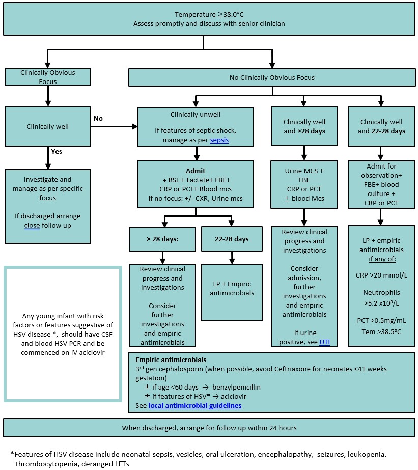
- Febrile infants 22–28 days (corrected age): are slightly lower risk. If well-appearing with low-risk inflammatory markers, they still require admission, but lumbar puncture and empiric antibiotics may be deferred
- Febrile infants <3 months (corrected age): need a lower threshold for investigation and treatment than older children. They may be discharged after observation if a source is identified, they remain clinically well, inflammatory markers
are reassuring, and follow-up is arranged
- In an unwell child, viral symptoms or a positive respiratory viral test should not solely be assumed to explain fever, as serious bacterial infection may coexist
Background
- Fever is defined as a body temperature of 38.0C
- Most febrile illnesses in children are caused by viral infections, however careful assessment is required to exclude a serious bacterial infection (SBI)
- Urinary tract infection is the most common SBI, followed by pneumonia. Other important SBIs in children include sepsis,
meningitis, bone and joint infections, skin and soft tissue infections and mastoiditis
- Unvaccinated children with fever who look unwell are at significantly greater risk of SBI and should be treated as sepsis
- The severity of illness cannot be predicted by the degree of fever, or its response to antipyretics. The child's age, appearance and behaviour are better indicators of serious illness (see Traffic light system for identifying risk of serious illness
below)
- Children with fever for ≥4 days should be assessed for Kawasaki disease / PIMS-TS.
For children with fever for >7 days without a source, refer to Prolonged fever
Assessment
History
- Fever timeline
- Localising symptoms: cough, headache, photophobia, diarrhoea, vomiting, abdominal pain, urinary changes, rash, musculoskeletal pain or reduced limb movement
- High-risk groups: neonates, unvaccinated children, immunocompromised children (secondary to a medical condition or immunosuppressive medications such as steroids, chemotherapy), children with central venous access devices, Aboriginal
and Torres Strait Islander children
- Immunisation history
- Current or recent antibiotic use
- Sick contacts
- Oral intake, urine output
- Overseas travel (See Fever in the recently returned traveller)
Note: Fever should not be attributed solely to teething or recent vaccination in an unwell child, and SBI should be considered
Red flag features
- High level of parental concern
- Representation within 48 hours
- Clinical deterioration despite previous treatment
- Recent surgery or burns
- Unwell children from high-risk group (see above)
Examination
All children should have a thorough clinical examination to identify a source of infection and assess for concerning features suggestive of SBI
Traffic light system for identifying risk of serious illness
|
Clinically well appearing child |
Clinically unwell appearing child |
| Category |
Low risk (no unwell features) |
Intermediate risk |
High risk |
| Colour |
Normal colour |
Pallor reported by parent/ carer |
Pallor, mottled
Cyanosed |
| Activity |
Normal response to social cues
Content, smiles
Stays awake or awakens easily
Strong cry or not crying
Older child: Normal activity
|
Poor smile/ response to social cues
Decreased activity
Wakes only with prolonged stimulation
Older child: Lethargic |
No response to social cues
Does not wake or if roused does not stay awake
Weak, high-pitched or continuous cry
Older child: altered mental state or behaviour |
| Breathing |
Normal effort of breathing |
Tachypnoea (Range for physiological variables)
SpO2
<92%
Increased work of breathing
Crepitations on auscultation |
Grunting
Apnoea
SpO2
<90% |
| Circulation / Hydration |
Normal skin and eyes
Moist mucous membranes |
Tachycardia (Range for physiological variables)
CRT ≥ 3 seconds
Reduced urine output
Signs
of dehydration |
Persistent, unexplained tachycardia
Hypotension
Cool peripheries |
| Other |
No Intermediate or high-risk features
Not from the high-risk groups (see history) |
Rigors
Swelling of a limb or joint
Non-weight bearing limb/ not using an extremity
Hypothermia (T
<36 °C)
Parent or carer concern that child is behaving differently from usual |
Non-blanching petechial or purpuric rash
Status epilepticus, focal seizures, focal neurological signs, neck stiffness, bulging fontanelle
Appears unwell to a healthcare professional
High level of parental concern that
child is deteriorating |
| Adapted from: Suspected sepsis in under 16s NICE Guideline 2025 |
Investigation and management
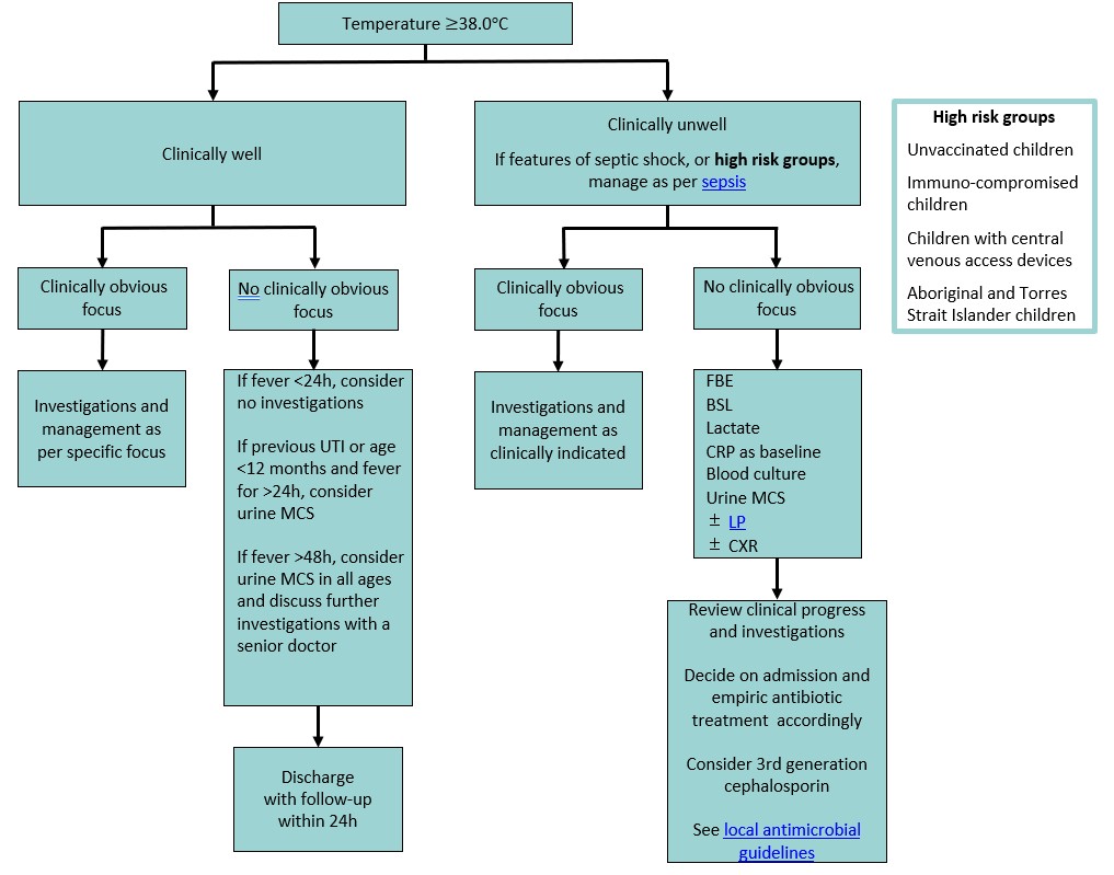
- Guided by age, clinical appearance (see table above), and whether a source of infection is identified
- Febrile children with high-risk features, or multiple intermediate-risk features, should be reviewed by a senior clinician. Higher risk warrants a lower threshold to treat as suspected sepsis,
regardless of fever degree
- In intermediate risk children, it is preferable to complete investigations for an infective focus before starting antibiotics
- In remote settings where limited access to investigation might delay care, it is safer to start antibiotics empirically while working the child up for the cause of fever
- Well-appearing neonates with only suspected, tactile or infrared-recorded fever may be suitable for a period of close observation before proceeding to a full septic work-up
- UTI is the leading cause of SBI and is particularly common among non-toilet-trained children. Urine collection and testing should be performed if there is no clinically obvious focus (See Additional notes below)
- When blood cultures are indicated, ensure adequate volume is collected (see Additional notes below)
Antimicrobial recommendations may vary according to local antimicrobial susceptibility patterns; please refer to local guidelines
Herpes simplex virus (HSV)
- Disseminated HSV presents most commonly in the first 6 weeks of life
- Clinical features of HSV disease include sepsis presentation, vesicular rash (absent in 30%), seizures, encephalopathy, hypothermia, mucous membrane ulceration
- Laboratory features of disseminated HSV include leukopenia, thrombocytopenia, deranged LFTs, coagulopathy
- A negative maternal history of genital HSV does not rule out neonatal infection, as most affected mothers are unaware of their status until the infant's diagnosis
- If concerned, perform HSV PCR testing on blood, CSF and skin lesion samples and commence treatment with aciclovir
Infants ≤21 days corrected age
- Assess promptly for signs of sepsis and discuss with senior clinician. See Recognition of the seriously unwell neonate and young infant
- FBE, CRP, blood culture, urine MCS by suprapubic aspirate (SPA). For other methods of urine collection: see Urine collection below, lumbar puncture (LP) for CSF studies (see Lumbar puncture),
± CXR
- Prompt treatment with empiric antibiotics
- If the infant is clinically unwell or investigations are delayed, start antibiotics and aciclovir immediately. Use the IV or intraosseous route, especially if unwell. Reserve the IM route for clinically well neonates with difficult
venous access
Infants 22 days - 3 months corrected age
Children >3 months corrected age
Consider consultation with local paediatric team when
- Unwell child or septic shock
- Infants
<28 days corrected age with fever
- High-risk child (see history)
- Prolonged fever of unclear cause
- Barriers to follow-up within 24 hours due to social or external factors (consider admission)
Consider transfer when
Child requires care above the level of comfort of the local hospital
For emergency advice and paediatric or neonatal ICU transfers, see Retrieval Services
Consider discharge when
- Infants ≤28 days: All infants are admitted. For infants who were well appearing at presentation, consider discharge when the blood culture is negative at 36 hours. Studies in infants under 90 days have found that over 96% of blood cultures
are positive within 36 hours
- Infants 29 days - 3 months of corrected age: well-appearing, investigations normal, reviewed by senior clinician, follow-up has been arranged within 24-48 hours with local GP, paediatric acute review, or virtual care clinic
- Children >3 months corrected age: well-appearing, follow up has been arranged
Always advise parents to return for review if the child is deteriorating or they are worried
Parent information
Fever in children
Additional notes
Recommended temperature measurement
- Axillary (<3 months): Placed over axillary artery for 3 minutes
- Tympanic temperature (>3 months)
- Skin/Infrared thermometers: unreliable, not recommended
- Rectal (neonates): Screen with axillary first, use rectal only if fever is still suspected
- When indicated, should be performed without delay and, ideally, before the administration of antibiotics
- Contraindications include impaired conscious state, haemodynamic instability, focal neurological signs and impaired coagulation. In this circumstance, treatment for meningitis/encephalitis should be commenced and an LP performed when the child
is stable and there are no contraindications
- SPA (see Suprapubic aspirate): Gold standard in children up to 6 months (low risk of contamination)
- In/out catheter: useful if the bladder is underfilled, such as after a failed clean catch or SPA (discard first few drops of urine if possible, to reduce contamination) (low-medium risk of contamination). See Indwelling urinary catheter - insertion and ongoing care
- Midstream urine (MSU): preferred method for toilet-trained children who can void on request (medium risk of contamination)
- Clean catch: appropriate for pre-continent children who cannot void on request, but are not seriously unwell (medium risk of contamination)
- Bag urine: not recommended for culture due to high false positive rates (high risk of contamination)
- Specify method of collection on pathology request
Note: The perineal/genital area should be cleaned with saline-soaked gauze for 10 seconds before collecting midstream or clean catch urine
Blood culture
- Blood cultures should be taken using an aseptic technique
- The sensitivity of blood cultures improves with larger blood volumes, although there is an upper limit to the optimal blood-to-broth ratio
- Ideal volumes for blood culture collection:
- Neonates: target volume 1 mL, paediatric specific bottle (minimum 0.5 mL)
- Children
<20 kg: 2-4 mL, paediatric specific bottle
- Children >20 kg: 5-10 mL x2, in aerobic and anaerobic bottles
- Inoculate aerobic bottle preferentially, eg if inadequate volume for both bottles (ie
<10 mL)
- If inadequate volume for aerobic bottle, ie
<5 mL, use paediatric specific bottle
Last updated October 2026