What is a cerebral aneurysm?
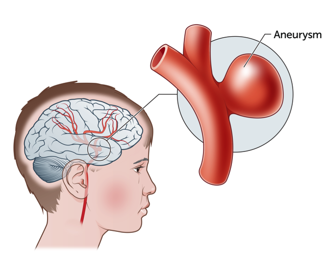
An aneurysm is a ballooned, weak area in the wall of an artery (blood vessel) supplying the brain. Brain aneurysms in children may be different to brain aneurysms in adults. Typically, aneurysms in children have no symptoms until they burst (rupture). A ruptured cerebral aneurysm can cause bleeding into the space around the brain, this is called a subarachnoid haemorrhage. This can extend within the brain and cause a blockage of the normal fluid in the brain or put pressure on the brain.

Signs and symptoms of a cerebral aneurysm
Unfortunately, the majority of patients with cerebral aneurysms don’t know they have one because the aneurysm typically causes no symptoms, until it bursts. A ruptured cerebral aneurysm will bleed over the surface of the brain and may extend into the brain, causing a haemorrhagic stroke.
What causes a cerebral aneurysm?
As children do not usually have any risk factors (such as high blood pressure, long-term excessive alcohol intake or high cholesterol) that increase their risk of cerebral aneurysms, often the cause in children is not understood. Researchers are working to learn more about the potential causes of aneurysms in children such as whether there are any genetic changes that may make this more likely.
How are cerebral aneurysms diagnosed?
A cerebral aneurysm may be diagnosed by a CT scan, or an MRI or MRA scan.
MRI/MRA scan: An MRI (Magnetic Resonance Imaging) machine or scanner uses a powerful magnet to take very clear and detailed pictures of the body. It is useful for looking at many parts of the body and often gives extra information to plain X-rays, ultrasounds or CT scans. During an MRI scan, the part of the body being scanned will have images taken from several different angles. There is no ionizing radiation (e.g. X-rays) used in an MRI and the magnetic field used in MRI is safe even during the second half of pregnancy.
In addition to the MRI, an MRA may also be completed. An MRA or magnetic resonance angiogram specifically looks at the blood vessels in the brain and neck and those around the stroke. The MRA and MRI are completed at the same time during the same scan.
CT scan: a CT scan or computed tomography imaging is a type of X-ray. A CT scan is not painful. The CT scanner is a big open doughnut-shaped machine. Your child will lie down on a table, which moves through the middle of the machine at least twice during the scan. The CT scanner takes all of its pictures as the table is moving.
Your child will need to remain very still for the pictures and sometimes hold their breath; usually this is for less than 10 seconds. Generally, the CT scan study takes about 10–15 minutes in total. With and injection of CT contrast (dye) a CT angiogram may be performed to show the blood vessels.
Cerebral angiogram: is an x-ray test where a dye (contrast) is injected directly into an artery through the groin whilst your child is under anaesthetic. This test gives detailed pictures of the arteries and veins (blood vessels) of the brain. This test is performed to look for abnormalities in blood vessels. Once the angiogram is completed, your child will be required to remain lying flat for four hours to ensure there is no bleeding from the artery in the groin.
All of these tests provide different information and your child may need a combination of these to plan the optimal treatment for them.
Treatment for Cerebral Aneurysms
A burst or ruptured aneurysm may require emergency surgery to drain cerebro-spinal fluid (CSF) (CSF is a clear and colourless fluids that is found in the brain and spinal cord) from your child’s brain, to relieve pressure. A drain, known as an External Ventricular Device (EVD), may be required, and drains CSF from the fluid pathways in the brain to an external collection bag.
Once an aneurysm has bled, the aneurysm needs urgent treatment to stop it from bleeding again. There are two main treatments for aneurysms. One treatment involves a surgical operation, and the other involves blocking the aneurysm through the blood vessels. The shape and location of each aneurysm determines which technique is the best option to treat them. The treating team caring for your child will discuss these options and explain what is recommended for your child.
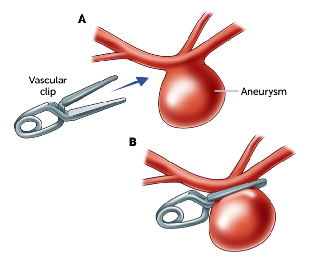
The surgical operation is referred to as ‘clipping’ an aneurysm which involves placing a titanium clip across the neck of the aneurysm, to stop blood flow into the aneurysm. Clipping of an aneurysm is performed under general anaesthetic. A small opening is made in the skull by a neurosurgeon, this is called a craniotomy. Through the small opening, the brain is retracted (moved back) and the aneurysm is located. The small clip is then placed across the base, or the neck of the aneurysm to block the blood flow from entering the aneurysm whilst allowing normal blood flow to continue in the main artery. The clips are made from titanium and remain on the artery forever.

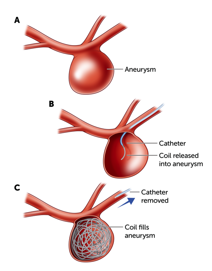
The other method of treatment is “coiling” an aneurysm. This involves filling the aneurysm with tiny platinum coils that cause the blood inside the aneurysm to clot. Coiling is performed with tiny instruments (a catheter) that are guided along the blood vessels from an artery in the groin. Complex aneurysms may also be managed by the placement of a stent. A stent is a tiny tube that is inserted into a blocked passageway to recreate a normal blood vessel channel. A stent stops the opening to the aneurysm.

Recovery following clipping of a cerebral aneurysm
If the aneurysm has not burst, the average stay in hospital is five days, with the overall recovery time typically taking four to six weeks. During this time, children will have restrictions on certain activities, such as contact sports. Your child’s doctor will advise on a gradual return to activities such as school, and sports following the surgery.
Recovery following coiling of a cerebral aneurysm
Immediately after the procedure is complete, your child needs to lay flat for approximately six hours to avoid bleeding in the groin where the catheter was inserted. Patients are then able to move around the room after a few hours of strict bed rest. Coiling of aneurysms does not require open surgery to the brain, and therefore if the aneurysm has not burst recovery is quicker. Your child’s doctor will advise on a gradual return to activities such as school, and sports following the surgery.
After an aneurysm has ruptured
It is important to understand that after an aneurysm has ruptured your child will need to recover from the bleed as well as having treatment of the aneurysm which prevents further bleeds.
As a result of the bleed, your child may experience problems with:
Vasospasm: a constriction or narrowing of the blood vessels in response to the bleed.
Hydrocephalus: a build-up of the cerebro-spinal fluid (CSF) within the brain
Ischaemic Stroke: which may occur as a result of vasospasm or due to the build-up of pressure on the brain, obstructing the flow of blood.
Your child’s treating medical team will be watching for symptoms such as:
- Altered level of consciousness
- weakness on one side of the face and/or body
- difficulty speaking
- confusion
- neck stiffness (less common)
- fever (less common)
In order to closely monitor your child’s progress following the bleed, your child might need to have further scans.
Follow-up
Your child should have regular surveillance imaging and follow-up appointments with the Neurosurgery department at their hospital.
Key points to remember
- An aneurysm is a ballooned, weak area in the wall of an artery (blood vessel) supplying the brain.
- Aneurysms in children have no symptoms until they burst (rupture) causing bleeding into the space around the brain.
- Cerebral aneurysms can be diagnosed on a CT scan, MRI or cerebral angiogram.
- Treatment for cerebral aneurysms include clipping or coiling of the aneurysm.
- Recovery following the rupture of an aneurysm can take many weeks.
For more information
Common questions our doctors are asked
When can my child return to school?
The duration that each child takes to recover from treatment for a cerebral aneurysm varies for each individual and is dependent upon what form of treatment was required and whether the aneurysm was un-ruptured or had bled. If your child suffered an intracranial bleed, their recovery may be much longer and they may not be able to return to school for several months and they may require a period of outpatient rehabilitation. Return to school may be possible sooner if your child has had treatment for an un-ruptured aneurysm.
Can my child resume normal sporting activities after clipping or coiling of a cerebral aneurysm?
Normal sporting activities can be resumed after clipping or coiling of a cerebral aneurysm but this should only be resumed after consultation with your treating doctor, and is likely to be following several weeks of recovery. Sports should be introduced gradually, with non-contact sports permitted first and contact sports only after full recovery including adequate healing of a craniotomy site (usually three months).
Can my child have an MRI or go through metal detectors after clipping or coiling of an aneurysm?
All of the modern clips, coils and stents are safe for clinical MRI ( not high strength scans) and will not activate metal detectors. However all patients undergoing an MRI scan must complete a safety questionnaire to determine if they have any implants. This must be completed as not all patients with implants can have an MRI scan. Each implant has a unique reference and provided this information is available prior to your appointment. The MRI team can determine whether an MRI scan is safe for your child. If the information is not available, the decision to determine if your child can safely have a scan is complex and may lead to the scan being cancelled so it’s very important that you bring any information you have about the implant to your appointment. Some implants may be able to be scanned on a 1.5 T rather than a stronger 3T magnet. Metallic artefact will be created in the area around the implants, which may obscure some anatomical detail.
The coils used to treat a patient's vascular malformation will be documented in the patient record and will need to be checked to ensure MRI safety. Aneurysm clips and endovascular coils will not be detected when your child walks through a metal detector but may be detected by high strength hand held devices.
Developed by The Royal Children's Hospital Neurology and Neurosurgery departments. We acknowledge the input of RCH consumers and carers.
Reviewed April 2020
Kids Health Info is supported by The Royal Children’s Hospital Foundation. To donate, visit
www.rchfoundation.org.au.