Introduction
Surgical drains are tubes placed near surgical incisions in the post-operative patient, to remove pus, blood or other fluid, preventing it from accumulating in the body. The type of drainage system inserted is based on the needs of patient, type of surgery, type of wound, amount of drainage expected and surgeon preference.
Aim
This guideline is designed to ensure a standard approach to care and management of surgical drains (as listed below) through evidence-based practice.
Note:
Types of drains
Assessment
Initial
- Surgical drains should be assessed 1-4 hourly throughout the shift
- Assess drain insertion site for signs of fluid or air leakage, redness or irritation to the skin.
- Document site condition and notify treating team and AUM if any concerns.
- Assess if the drain is maintaining suction
- Assess securement type and document on LDAs.
- Assess patency of drain. Ensure drain is located below the insertion site and free from kinks or knots.
- Document amount, output appearance, type of fluid in drain bottle/receptacle and drain status on LDAs.
Ongoing
- Monitor for infection. Signs of infection include: redness, tenderness at the drain site, warmth at site, increased ooze, or a change in collection fluid to purulent, or if the patient is febrile.
- Drain patency and insertion site should be observed at the beginning of your shift and before and after moving a patient. A kinked, disconnected, dislodged or blocked drain tube can lead to formation of haematoma, increased pain and risk of infection.
- Drainage needs to be documented at a minimum 4 hourly and more frequently if output is high. This needs to be documented in flowsheets in the sections “Output in previous hours” and “Chamber reading” so an accurate fluid balance is maintained.
- Suction needs to be assessed throughout the shift. Suction will no longer be maintained once the drain becomes full. This drain will need to be emptied, changed or suction reapplied.
- Discuss removal plan with treating team. Drains should be removed as soon as practicable, the longer a drain remains in situ, the higher risk of infection or development of granulation tissue. This can cause pain and trauma upon removal.
- Pain should be assessed whilst the drain is in situ. Appropriate analgesia should be provided when necessary. Please refer to the
pain assessment and management guideline for more information.
Investigations
If infection is suspected, notify the treating medical team as a swab of the insertion site may be required.
Education
- Educate the patient/parent to ensure the drain is below the site of insertion but not pulling on the patient.
- Educate the patient/parent that there is a risk of dislodgement therefore requiring increased care when moving.
- The patient should be aware that moving whilst drain is in situ will cause some pain, but this can be minimised with regular analgesia.
- The patient should be encouraged to mobilise with supervision when appropriate.
Troubleshooting
Reinstating Suction
This is dependent on the type of drain. As fluid collects in the drain, the unit either expands or becomes full and negative pressure is lost. The drain is then ineffective and needs to be emptied or changed to reinstate suction.
N.B. Suction is required
unless specifically stated otherwise by treating team.
Redivac: To signal that suction is being maintained the green vacuum indicator on top of the drain should appear pressed down. If the green vacuum indicator is fully expanded, then the redivac needs to be changed. Ensure “standard aseptic technique” is ulitised when the drain is changed. If suction is unable to be maintained, the treating team should be notified. |
 Example of Redivac drain that is not maintaining suction |
Jackson Pratt: The bulb of the drain will appear like it has been squeezed to demonstrate that suction is being maintained. If the bulb appears expanded, kink the tube above the bulb, pull the output. Then squeeze the bulb and insert the plug back into the drain.
|
 Example of how to reinstate suction on Jackson Pratt |
Bellovac: If there has been no drainage:
ensure bellovac is below wound and gently shake sideways and give bellows 2 quick squeezes to start flow without vacuuming.
Mini-vacuum
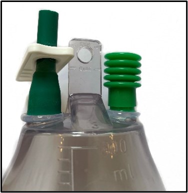
drain: The bellows can be twisted off from the cap and squeezed together to increase vacuum.
Emptying
Redivac: drain cannot be emptied. Once the drain is full document output into flowsheets and change drain container.
Jackson-Pratt: kink the tube above the bulb and pull the plug out. Empty the contents, measure and document output. Then compress or squeeze the bulb and insert the plug back in to close bulb.
Bellovac: close the clamp above the bellows, ensure the clamp below the bellows is open. Compress the bellows fully, this can be done slowly and in stages. The bellows will not re-expand due to the one way valve. Fluid from the bellows should drain into the collection bag. Re-open the clamp above the bellows.
Mini-vacuum drain: only empty if drain is full. Can be twisted off from the cap to empty output. Output should be minimal and emptying this drain is not usually indicated. If there is a large amount of output, notify the treating team.
Penrose and Pigtail: gauze or contents in drainage bag should be weighed and documented in flowsheets
Moving a patient with a drain tube
Assess the patient including all drains and attachment sites prior to mobilising. Ensuring drains are secured and will not dislodge/pull on patient.
When appropriate, patient mobilisation with a drain should be encouraged to reduce risk of DVT and enhance recovery.
Reassess drains post mobilising to ensure dislodgement of drains has not occurred.
At all times, ensure drainage tube is not entangled with other leads (IV tubing, O2 leads, etc.) as this could lead to inadvertent removal of the tube.
Leakage
If leakage occurs at a surgical drain site, please notify the AUM and treating team and consider the following:
- Redress or retaping the surgical drain dressing (preferably with an occlusive dressing) using standard aseptic technique.
- Placing a Coloplast™ drainage bag (2245) over the surgical drain tubing.
- Consider taking a clinical image on EPIC if deemed necessary.
- Review the
wound care nursing guideline for further information.
- Refer patient to Stomal Therapy for further input if necessary.
Blockage
If drainage is minimal, ensure the drain is not blocked. If blocked, notify the treating team and AUM.
Inadvertent removal/Drain dislodgement
- If the drain is suspected to have moved position, the drain should be secured and the treating team notified.
- In the event a drain has been removed or dislodged, a sterile dressing should be applied and the treating team notified immediately.
- If the drain is suspected to have receded into the patient, the treating team should be notified and imaging (x-ray, etc.) should be performed.
Link to Policy & Procedure: Surgical Wounds – Procedure for Missing/Non Intact Drains
Removal
Things to consider prior:
- Discuss and plan for procedural pain management and non-pharmalogical interventions to minimise pain and distress throughout procedure, assess analgesic requirements first and then consider the need for procedural sedation; please refer to the
procedural sedation ward and ambulatory areas at RCH procedure for more information. If using analgesia ensure it is given 30-45 minutes prior to procedure to ensure it has taken peak effect. Please refer to the
procedural pain management guideline for more information.
- The technique for the following is ‘standard aseptic technique’ for a simple dressing” If the dressing is complex for example when wounds are near the drain or the patient is non-complaint, the procedure can be done using “surgical aseptic technique.” Please refer to the relevant policies for more information
(
Aseptic-Technique)
 |
ALERT:
Pigtail drains must be uncoiled prior to removal, failure to uncoil a pigtail drain can cause severe pain and/or tissue damage. To uncoil the pigtail drain the catheter/string should be cut to release the string that creates the pigtail coil. Non-locking pigtails do not need to be uncoiled.
|
1. Ensure plan for removal of drain tube is discussed with and ordered by the treating team in the patient’s progress notes on EMR.
2. Inform patient/parent about removal process and possible associated pain, administer pain relief.
3. Ensure drain is taken off suction:
- Redivac: ensure both clamps are clamped
- Jackson Pratt: pull plug out of bulb and ensure the bulb is fully expanded
- Bellovac: slide the clamp above the bellows up the tubing to the point just below the connection to the catheter and close it off. Needs to be un-clamped 30 minutes prior to removal to allow the pressure in the catheter to dissipate
- Mini-vacuum: ensure bellows appears expanded
4. Clean work surface with detergent and prepare waste bag.
5. Perform hand hygiene.
6. Identify and collect all equipment for procedure.
7. Perform hand hygiene.
8. Open aseptic field (small general or large critical) and peel open any additional sterile equipment and drop onto field.
9. Perform hand hygiene.
10. Prepare patient, use gloves if removing dressings.
11. Remove gloves if worn and perform hand hygiene, if using surgical aseptic technique don sterile gloves.
12. Perform procedure ensuring all key parts are protected.
13. Sterile items are used once and not returned to the aseptic field; waste is disposed into waste bag.
14. Clean around the site with normal saline and remove any sutures.
15. Rotate tubing from side to side gently to loosen, then remove the drain using a smooth, but fast, continuous traction.
16. Immediately apply occlusive dressing with gentle pressure until bleeding or oozing stops.
17. Inspect drain for intactness.
18. If required, cut the tip of the tube for cultures.
19. Remove gloves and perform hand hygiene.
20. Clean work surface, dispose of waste and perform hand hygiene.
21. Document removal of drain and that it is intact/not intact in LDA’s and progress notes as well as amount of drainage in the flowsheets. If drain is not intact report to treating team and keep drain for further inspection.
Unable to remove surgical drain
If there is resistance and no movement of the drain tube despite gentle side-to-side rotation and a firm pull do not proceed further and notify the treating team/surgeon.
There should be no excessive force when pulling the drain tube, doing so can lead to serious complications such as drain tube fractures or internal tissue damage.
Drain Tube Fractures
If the tube fractures during drain removal and remnants of the tubing is left within the patient contact the treating team immediately.
The surgical fellow should order an immediate X-ray of the drain tube site.
The patient should be prepared for theatre, inform the parents and consider the need to keep the child nil by mouth in anticipation for surgical removal of the remaining drain tube.
The whole drain unit should be kept in the patient’s room until surgical review and will need to be kept for collection to enable quality review.
The piece of drain tubing that remains in the patient will also be kept once surgically removed to allow for appropriate follow up of the incidents cause.
A VHIMS must be completed by the nurse delegated to remove the drain.
For Theatre Staff involved in the surgical removal retained drain tube:
In theatre, previous surgery is checked on EPIC regarding the LDA’s flowsheet of the drain that was inserted at that operation. NB. There can be multiple drains.
After removal of retained drain, instrumentation nurse to superficially clean visible blood /serous fluid off the retained drain.
Instrumentation nurse to measure length of retained drain before placing in a yellow top container. -Surgery is completed as planned.
Scout RN to record length of retained item on patient’s UR label on container.
Retained item's lot number and expiry that has been recorded in EPIC is to be transcribed onto the yellow top container with patient’s UR label.
Scout RN to record in EPIC retained items details. Also check LDA's flow sheet for removal of drain/line details and update in comments section that residual item has been removed and record length with date and time stamp.
Yellow top container to be kept together with the remaining drains until critical review process is completed and VHIMs documentation finalised.
Post op X-ray to be reviewed by surgeons and open disclosure to family to be undertaken by surgeons.
Link to Policy & Procedure: Surgical Wounds – Procedure for Missing/Non Intact Drains
Link to aseptic technique policy and procedure
Post removal
Monitor site for signs of infection, obtain swabs or samples if required.
Monitor and mark dressings to ensure minimal leakage, replace dressings as required to minimise risk of infection. Excessive leakage should be reported to AUM or surgeon.
Dressing should be removed when wound has healed (3-5 days).
Links
Please remember to read the
disclaimer.
The development of this nursing guideline was coordinated by Madeleine Lennox, RN, Specialist Clinics, and approved by the Nursing Clinical Effectiveness Committee. Updated November 2023.
Evidence Table
| Reference
(include title, author, journal title,
year of publication, volume and issue, pages) |
Evidence
level
(I-VII) |
Key
findings, outcomes or recommendations |
| Knowlton, M. C. (2015). Nurse's guide to surgical drain removal. Nursing, 45 (9), 59-61. doi: 10.1097/01.NURSE.0000470418.02063.ca. |
|
General overview of types of drains, removal of surgical drains, how long drains should stay in place before infection risk increases and complications of removal.
|
| Tanaka K, Kutamoto T, Nojiri K, Takeda K, Endo I. (2013) The effectiveness and appropriate management of abdominal drains in patients undergoing elective liver resection: a retrospective analysis and prospective case series. Surgery Today, 43(4), 372-80. Epub 2012 Jul 14. |
IV |
Ascending infections can be avoided by early drain tube removal
|
Bharathan R, Dexter S & Hanson M. (2009). Laparoscopic retrieval of retained Redivac drain fragment. Journal of Obstetrics and Gynaecology, 29(3), 263-264
|
VI |
Case study demonstrating the effectiveness of post drain removal xray to identify drain fragment, this then resulted in successful removal of fragment
|
| Durai R, Mownah A & Ng PCH (2009). Use of drains in surgery: a review. Journal of Perioperative Practice, 19(6), 180-186 |
V
|
A peer reviewed literature review of drains in surgical situations. Definition and uses of drains
|
| Havey R, Herriman E & O’Brien D. (2013) Guarding the gut: early mobility after abdominal surgery. Critical Care Nursing Quarterly, 36(1), 63-72. VII Importance of mobilizing with a drain, process on how to mobilize patient with a drain Durai R & Ng P. C. H. (2010). Surgical vacuum drains: Types, uses, and complications. AORN Journal, 91(2), 266-71; quiz 272-4. doi:http://dx.doi.org/10.1016/j.aorn.2009.09.024 Overview of drain management, including movement, removal and complications |
VII |
Overview of drain management, including movement, removal and complications
|
| Pagoti R, Arshad I & Schneider C. (2008). Stuck redivac drain: to open or not to open? A case report. Australian and New Zealand Journal of Obstetrics and Gynaecology, 48(2), 223-224. |
VII |
Case report detailing complications of a Redivac insertion
|
| Imm N (2015). Surgical Drains-Indications, Management and Removal. Available from: https://patient.info/doctor/surgical-drains-indications-management-and-removal. Accessed 04/10/2018. |
VII |
Based on research evidence and written by medical professionals including indications, management and removal of surgical drains
|
| Mediplast. (2019). Bellovac Closed Wound Drain System. Retrieved from https://www.mediplastresourcecentre.com.au/bellovac/ |
|
Product website that states management of bellovac drain |