Fracture Guideline IndexSee also: Radius /ulna shaft (diaphysis) fractures - Fracture clinics
- Summary
- How are they classified?
- How common are they and how do they occur?
- What do they look like - clinically?
- What radiological investigations should be ordered?
- What do they look like on x-ray?
- When is reduction (non-operative and operative) required?
- Do I need to refer to orthopaedics now?
- What is the usual ED management for this fracture?
- What follow-up is required?
- What advice should I give to parents?
- What are the potential complications associated with this injury?
1. Summary
Fracture Type
|
ED Management
|
Follow-up
|
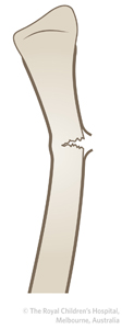
Greenstick
|
Refer to acceptable angulations
Closed reduction with immobilisation in above-elbow cast for 6 weeks. Three point moulding is required
|
Fracture clinic within 7 days with x-ray
|
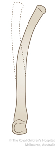
Plastic deformation
|
Plastic deformation is often unrecognised. Radiographic findings may be subtle. It is most commonly seen in the ulna. Refer to orthopaedics for advice. Usually requires general anaesthetic manipulation plaster (GAMP) due to prolonged force to correct deformity
|
Fracture clinic within 7 days with x-ray
|
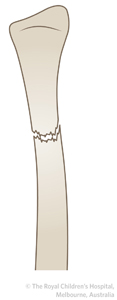
Complete fracture
|
Refer to acceptable angulations
Closed reduction with immobilisation in above-elbow cast for 6 weeks. Three point moulding is required
|
Fracture clinic within 7 days with x-ray
|
2. How are they classified?
Forearm shaft fractures can be classified by the following:
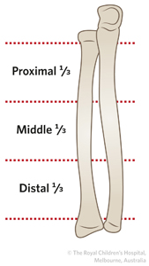
a) Fracture location: proximal, middle or distal third
|
|
b) Fracture pattern:
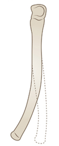
- Plastic deformation : bowing without disruption of cortex
See fracture education module for more information
|
|
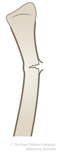
- Greenstick fracture (incomplete fracture): an incomplete fracture, in which only the convex side of the cortex is broken with bending of the bone
See fracture education module for more information
|
|
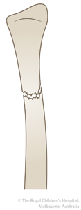
See fracture education module for more information
|
|
c) Bone involvement: single bone or both bones
|
|
3. How common are they and how do they occur?
The rate is approximately 1 in 100 children per year. Peak age of incidence is 12-14 years. They are the most common open fracture in the upper extremity for the paediatric population.
The most common mechanism is a fall onto an outstretched hand. Rotational forces through the forearm can cause the fractures of the radius and ulna to be at different levels. Direct impacts can cause an isolated fracture of one bone, more commonly the ulna.
4. What do they look like - clinically?
In displaced fractures, there is usually deformity, pain and tenderness directly over the fracture site and limited range of forearm rotation (supination and pronation).
There may be only subtle findings with plastic deformation and greenstick fractures.
5. What radiological investigations should be ordered?
✓
|
True anteroposterior(AP) and lateral views to include the wrist and elbow joint (whole forearm) should be ordered.
|
If the elbow and wrist are not adequately visualised, AP and lateral views of these joints should be obtained to eliminate Monteggia fracture-dislocation, supracondylar humeral fracture, lateral condyle fracture and Galeazzi fracture-dislocation.
6. What do they look like on x-ray?
Plastic deformation
!
|
Plastic deformation is often unrecognised. Radiographic findings may be subtle. It is most commonly seen in the ulna.
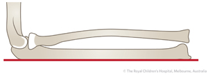
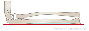
TIP: On x-ray, the normal ulna is straight and the normal radius is bowed.
|
|
Normal ulna with straight border (red line).
|
Plastic deformation of the ulna. Notice that the ulna border is not straight (shaded area).
|
|

Figure 1: Plastic deformation (red line) of one bone is usually associated with a fracture of the other forearm bone. It is best seen on the lateral view. In this case, there is also a dislocation of the radial head (Monteggia fracture-dislocation).
Greenstick fracture
Figure 2: AP and lateral x-ray of forearm showing greenstick fracture of radius and ulna in the middle third of a five year old. The radial greenstick fracture is more clearly seen on the lateral x-ray than the AP.
Complete fracture
Figure 3: 10 year old boy with transverse fracture of the diaphysis midshaft. There is also a greenstick fracture of the midshaft ulna.
| ! |
In single bone fractures,the proximal and distal radioulnar joints should be carefully inspected on x-ray. An isolated ulna fracture may be associated with dislocation of the radial head (Monteggia fracture-dislocation). These fractures should be referred to the nearest orthopaedic service on call.
An isolated radius fracture may be associated with dislocation of the distal radioulnar joint (Galeazzi fracture-dislocation or Galeazzi equivalent). These fractures should be referred to the nearest orthopaedic service on call.
|
7. When is reduction (non-operative and operative) required?
Table 1: Acceptable angulations for shaft fractures of the radius.
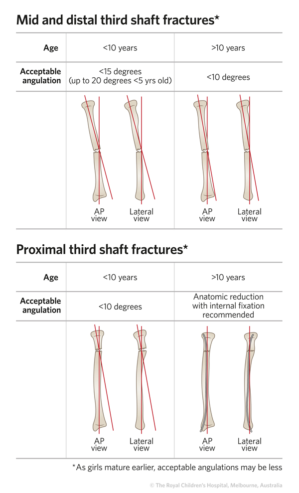
*Adapted from Price (2010) and Parikh et al (2010).
Up to 45 degrees of rotation is acceptable. However, as rotation is very difficult/impossible to quantify on x-rays. Any fracture with demonstrable rotation should be referred for an orthopaedic opinion.
Angles post-reduction should be within the same parameters for acceptable limits of alignment (see Table 1).
| ✓ |
If the forearm looks deformed clinically, the fracture will usually need a reduction. If the deformity can only be seen on x-ray, it may need a reduction.
Post reduction x-rays in the cast must be performed
|
8. Do I need to refer to orthopaedics now?
Indications for prompt consultation include:
- Open fractures
- Neurovascular injury with fracture
- Extreme swelling/compartment syndrome
- Unable to achieve or maintain reduction (including if ED is not experienced in fracture reduction, splinting or casting)
- Forearm fractures with elbow or wrist dislocations (Monteggia and Galeazzi)
- Ipsilateral upper extremity fracture
- Plastic deformation
9. What is the usual ED management for this fracture?
Table 1: ED management of radial shaft (diaphysis) fractures
Fracture type
|
Type of reduction
|
Immobilisation method & duration
|
Greenstick
|
Closed reduction
This fracture is suitable for a local anaesthetic, manipulation and plaster (LAMP)or procedural sedation in the ED, provided that there are appropriate resources and accredited personnel at your health service
|
Above-elbow cast for 6 weeks. Three point moulding, interosseous moulding and straight ulnar border is required. Elbow should be placed in 90 degrees flexion and forearm in midprone position. If the fracture is dorsally angulated, place wrist in slight flexion
|
Interosseous moulding
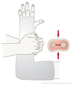
The cast should have a good interosseous mould, with an oval rather than a circular cross section, because this helps to maintain tension in the interosseous membrane
|
Plastic deformation
|
Refer to the nearest orthopaedic on call service for advice
Usually requires general anaesthetic manipulation plaster (GAMP) due to prolonged force to correct deformity
|
As above
|
Complete
|
Closed reduction with procedural sedation or GAMP
|
As above
|
10. What follow-up is required?
Patients should be seen in fracture clinic within 7 days for reassessment with x-ray.
11. What advice should I give to parents?
With proper treatment, most displaced fractures of the radius and ulna have an excellent outcome. Loss of forearm rotation can occur if the fracture does not heal within acceptable ranges of angulation. There is a very low risk of growth arrest in this injury.
For displaced fractures, the need for close follow-up should be emphasised due to the risk of loss of reduction.
12. What are the potential complications associated with this injury?
| ! |
A missed injury, especially in single bone fractures is a common complication.
With an isolated ulna fracture, check for injury to radiocapitellar joint (Monteggia fracture-dislocation).
With an isolated radius fracture, check for injury at the wrist (Galeazzi fracture-dislocation).
|
Other early complications include:
- Loss of reduction
- For displaced fractures of the diaphysis, up to one in four will lose position and will need a re-reduction. This risk is higher in complete fractures of both bones. Loss of position and the opportunity for re-reduction can only happen with appropriately timed follow-up
- Poor cast technique and residual angulation/displacement after the initial reduction are two major factors that can cause subsequent loss of alignment
- Compartment syndrome
- Compartment syndrome due to restriction by cast. The arm should be elevated post-casting and watched closely to assure that the cast is not too tight. Patients may need overnight observation
- Refracture
- Approximately one in twenty (5%) will have a refracture within 6 months of injury. The risk is higher immediately after cast removal
See fracture clinics for other potential complications.
References (ED setting)
Mehlman CT, Wall EJ. Injuries to the shafts of the radius and ulna. In Rockwood and Wilkins' Fractures in Children, 7th Ed. Beaty JH, Kasser JR (Eds). Lippincott Williams & Wilkins, Philadelphia 2010. p.347-404.
Parikh SN, Wells L, Mehlman CT, et al. Management of fractures in adolescents. J Bone Joint Surg Am 2010; 92(18): 2497-58.
Price CT, Flynn JM. Management of fractures. In Lovell and Winter's Pediatric Orthopaedics, 6th Ed, Vol 2. Morrissy RT, Weinstein SL (Eds). Lippincott, Philadelphia 2006. p.1464-71.
Price CT. Acceptable alignment of forearm fractures in children: Open reduction indications. J Pediat Ortho 2010; 30: S82-4.
Rang M, Stearns P, Chambers H. Radius and ulna. In Rang's Children's Fractures, 3rd Ed. Rang M, Pring ME, Wenger DR (Eds). Lippincott Williams & Wilkins, Philadelphia 2005. p.135-50.
Tredwell SJ, Van Peteghem K, Clough M. Pattern of forearm fractures in children. J Pediat Ortho 1984; 4(5): 604-8.
Feedback
Content developed by Victorian Paediatric Orthopaedic Network.
To provide feedback, please email rch.orthopaedics@rch.org.au