Fracture Guideline Index
See also:
Slipped upper femoral epiphysis (SUFE) - Fracture clinics
1. Summary
|
ED management
A child presenting with a chronic slipped upper femoral epiphysis (SUFE) will generally walk with an antalgic gait, out-toeing and some shortening of the affected limb. The child may complain of vague pain in the groin, thigh or knee.
A very reliable clinical sign of a chronic SUFE, even when mild, is obligatory external rotation of the leg during hip flexion.
Anteroposterior (AP) and frog lateral pelvis x-rays of both hips should be obtained.
All patients with a SUFE should be admitted under orthopaedics, as the management of SUFE is always surgical. Where the diagnosis is suspected, the patient should strictly be kept non weight-bearing in a bed or wheelchair (not crutches) until orthopaedic assessment takes place.
A guideline with a broader approach to the
limping child is also available. |
2. How are they classified?
A SUFE is characterised by the displacement of the capital femoral physis from the metaphysis. They can be classified according to:
- Ability to weight bear
- Stable - the patient is able to weight bear on the affected leg
- Unstable - the patient is unable to weight bear on the affected leg, even with crutches
- Duration of symptoms
- Acute - sudden onset of severe symptoms and inability to weight bear
- Chronic - gradual onset and progression of symptoms for more than 3 weeks, without sudden exacerbation. This is the most common presentation (85% of patients with SUFE)
- Acute on chronic - sudden exacerbation of symptoms due to acute displacement of a chronically slipped epiphysis
3. How common are they and how do they occur?
The aetiology of SUFE is unknown, but biomechanical and biochemical factors play an important role.
-SUFE is relatively common and occurs between 0.2 and 10 per 100,000 population.
-It is more common in boys (60%) than girls with the mean age at diagnosis being 13.5 years in boys and 12 years in girls.
-Approximately 50% of adolescents with SUFE are above the 95th percentile for weight.
-Studies have shown a risk of bilateral slips in 18 -50 % of patients.
4. What do they look like - clinically?
A child presenting with a chronic SUFE will generally walk with an antalgic gait, out-toeing and some shortening of the affected limb.
If the slip is acute and unstable, these children cannot walk. The child may complain of vague pain in the groin, thigh or knee. SUFE commonly presents with knee pain as the only presenting complaint.
A very reliable sign of a chronic SUFE, even when mild, is detection of obligatory external rotation during flexion of the hip. As the hip is flexed on the affected side, the thigh will automatically externally rotate and abduct .
It is common to see a child with months of symptoms having been treated for knee pain eventually diagnosed with a late SUFE |
5. What radiological investigations should be ordered?
In a suspected acute SUFE, order a standard AP pelvis X-ray, and a cross-table lateral x-ray, but NOT a frog-leg lateral xray as moving to this position may disrupt the vascular supply to the head of femur.
Where chronic SUFE is suspected and the patient has been bearing weight right up until presentation, it is useful to obtain a frog-leg lateral as well as an AP pelvis: again, this should NOT be ordered where an acute slip is suspected.
6. What do they look like on x-ray?
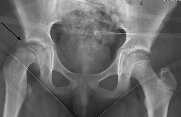
Figure 1: High Grade R SUFE
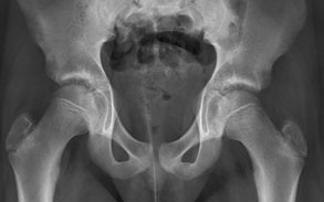
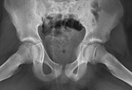
Figure 2 and 3: AP and frog leg views of a left sided SUFE
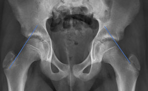
Figure 4: AP view of the same patient, showing Klein’s lines. On the normal right side, a line drawn along the neck of the femur shows a portion of the head of the femur above the line. On the affected left side, the head of the femur does not appear above the line.
7. Do I need to refer to orthopaedics now?
All patients with a SUFE or concern for a SUFE need urgent orthopaedic assessment.
8. What is the usual ED management for this injury?
All patients with a SUFE need surgical stabilisation.
The patient needs to be kept non-weight bearing (bed or wheelchair only - no crutches) and admitted for surgical treatment.
9. What advice should I give to parents?
Printed information is available and should be provided to parents.
If the child does have a SUFE, he or she will need surgery to stabilise the hip and must be admitted to a hospital that can manage the condition.
Some patients with SUFE are at risk of future slipped epiphysis of the contralateral painless hip. The orthopaedics team will consider whether prophylactic fixation of the contralateral normal hip is advisable and discuss this with the family.
Some patients presenting with SUFE carry risks of long term morbidity for their underlying medical conditions: an opportunity exists to refer them for weight management and lifestyle modification as appropriate.
10. What are the potential complications associated with this injury?
- Osteonecrosis - the risk is up to 50% in an unstable SUFE, even with treatment.
- Chondrolysis - this can result from the process of the SUFE itself, but more commonly it is from unrecognised screw/pin penetration from surgical stabilisation. The overall incidence of this is approximately 7%.
- Osteoarthritis - patients with a moderate or severe SUFE have higher risk of early degenerative joint disease.
- Impingement - patients with a severe SUFE have a risk of deformity through the femoral neck when the SUFE is stabilised and healed. This can cause femoral acetabular impingement, and may require further surgical treatment to correct this.
See fracture clinics for other potential complications.
11. References (ED setting)
Aronsson DD, Loder RT, Breur GJ, et al. Slipped capital femoral epiphysis: current concepts. J Am Acad Ortho Surg 2006; 14: 666-79.
Weigall P, Vladusic S, Torode I. Slipped upper femoral epiphysis in children: delays in diagnosis. Aust Fam Physician 2010; 39(3): 151-3.
Kay RM. Slipped femoral capital epiphysis. In Lovell and Winter's Pediatric Orthopaedics, 6th Ed, Vol 2. Morrissy RT, Weinstein SL (Eds). Lippincott, Philadelphia 2006. P.1085-124.
Mathew S. Natural History of Slipped Capital Femoral Epiphysis. J Pediatr Orthop 2019;39:S23-S27
Last updated December 2020