See also
Trauma - the primary survey
Head injury
Thoracic elevation device
Key points
- Cervical spine injury (CSI) is uncommon in children
- Maintain spinal precautions until cervical spine has been cleared
- Most cervical spines can be cleared clinically. A senior clinician must assist in the assessment of children with persistent symptoms or those who are unable to communicate
- The goal is to minimise movement. A soft collar may be used if tolerated until CSI is excluded. Hard collars are not recommended
Background
This guideline is for cervical spine assessment only
- This guideline addresses the ED assessment of possible CSI in children following blunt trauma. It does not cover the management of confirmed CSI
- Cervical spine assessment in children can be challenging. Assessment should be undertaken as soon as it is safe to do so with the aim of identifying CSI while avoiding unnecessary immobilisation and imaging
- The Paediatric Emergency Care Applied Research Network (PECARN) CSI clinical prediction rule and imaging algorithm has been derived and validated to support clinical clearance and guide imaging decisions after blunt trauma
- Common injury patterns differ in younger vs older age groups. Under 8 years of age, higher spinal cord injuries (C1-3) are more common, as opposed to predominantly lower cord injuries >8 years of age
Spinal precautions
- Minimise movement of the potentially injured spine
- Ideally the spine should be kept in a neutral and in-line position
- All children with suspected CSI should have spinal precautions maintained until the cervical spine is cleared (see Management)
- Particular care should be taken to maintain spinal precautions during moments of risk (see Appendix 2)
Assessment
History
Mechanism of injury informs clinical suspicion and the need for careful assessment. The presence of a high-risk mechanism should prompt senior review and ongoing reassessment. Persistent symptoms including pain, paraesthesia and/or weakness should prompt
further assessment and consideration of imaging
Risk factors for significant CSI in children
- Axial load to the head (diving, trampoline, falling from height)
- Forced neck hyperflexion (includes low velocity mechanism with high force eg rugby scrum collapse)
- Improper restraint in a motor vehicle collision (MVC)
- Passenger in an MVC at >60 km/hr, head on collision, rollover, ejection from any vehicle or any accident with a passenger death
- Pedestrian, bicycle or non-motorised vehicle struck by a motor vehicle
- Fall >3 metres (or twice the child’s height)
- Kicked by or fall from a horse
- Substantial head or torso injury (including the abdomen, flanks, back, spine, pelvis and clavicles)
- Clothesline injury and hanging
Conditions known to predispose to CSI (see full list Appendix 3)
- Previous CSI or injury
- Trisomy 21
- Osteogenesis imperfecta, achondroplasia
- Other rheumatological, congenital, genetic or metabolic conditions
Examination
Minimise movement of the potentially injured cervical spine, until it has been adequately assessed, and a targeted neurological examination performed
For reliable clinical examination, the child must be
- alert and cooperative
- developmentally able to participate in the examination
- not intoxicated
- not significantly sedated
- have sufficient analgesia for distracting injuries
Examining the cervical spine
Prior to palpation, ask the child if they have symptoms using age-appropriate questions
- Provide reassurance
- Observe the child including position, movement, distress
- Whilst minimising neck movement, gently palpate the posterior midline of the neck. Feel from the nuchal ridge to the 1st thoracic vertebra. The most prominent spinal process arises from the C7 vertebra.
- Repeat the process lateral to the midline on both sides
- Tenderness may indicate vertebral or soft tissue injury
- If there is no significant neck pain or tenderness and no abnormal neurology, assess the active range of movement of the neck by asking the child to slowly rotate their head to each side, place their chin to chest and look up. Children should be able
to move greater than 45 degrees to each side. Stop immediately if this causes pain or paraesthesia and minimise movement of the cervical spine
Children who are unable to communicate
- Should be assessed with assistance from a senior clinician
- Assessment is performed when the child is awake and alert by allowing the child to freely move their neck whilst they are observed carefully for restricted movement(s) or torticollis
- If the child demonstrates pain free and normal neck movement, and has no significant other injuries or neurological concerns, the cervical spine can be cleared
Red flags
- Traumatic torticollis
- Acquired twisting of the neck caused by injury to muscles, ligaments or vertebrae following trauma. Can present as an abnormal head position/tilt with reduced range of motion due to pain
- Child uses hand to support head or neck (this may be the only sign of an atlanto-occipital dislocation)
- Abnormal or asymmetrical neurology: focal motor or sensory deficit or severe headache
- GCS <15
- Child complains of neck pain or cervical spine tenderness
- Restricted neck movement
- Significant head, chest, abdominal, pelvic or other spinal injuries (ie those that require admission, investigations or surgery)
- Unexplained refractory hypotension (as a feature of neurogenic shock)
Investigations
PECARN risk stratification to guide imaging
High-risk features (CT)
- GCS 3-8 or AVPU = U
- Abnormal airway/breathing/circulation
- Focal neurological deficit
Intermediate-risk features (consider x-ray first line)
- Altered mental status (GCS 9-14 or AVPU V/P) or other symptoms of mental status change
- Self-reported neck pain or midline tenderness
- Substantial head or torso injury
Low-risk features (may be clinically cleared)
Imaging
CT scans
CT cervical spine should be considered first line for children with PECARN high-risk features. CT may also be considered when
- x-ray is abnormal or inconclusive
- there is persistent objective neurological abnormality of weakness or sensory loss
- the child is undergoing CT of other body regions; inclusion of the cervical spine may be considered following senior clinician review
X-ray
In PECARN intermediate-risk children, plain radiography is recommended as first-line imaging where feasible. However, senior clinician judgement, local resources and child specific factors including overall clinical picture should be considered in first
line imaging decisions
- ≤5 years old: AP and lateral X-rays only
- ≥6 years old: AP, lateral and odontoid X-rays are required
- Lateral X-rays should include views of the vertebral column from the occiput to T1 vertebra (this may require shoulder traction)
- Flexion and extension views are NOT recommended
If imaging findings (CT or X-ray) are inconclusive or the child has persistent symptoms or clinical signs (regardless of imaging findings), cervical spine precautions should be continued, and MRI/specialist consultation considered
Management
Restriction of cervical spine movement
- Maintain spinal precautions until CSI can be excluded. Expedite definitive imaging and senior review
- A soft collar is often useful to minimise movement (See appendix: instructions for soft collar). Also label with “C-spine not clear” until CSI has been excluded. However, do not reposition the neck for the following children who should
be left in a position of preference
- Children who use their hands to support the head
- Children with traumatic torticollis
- Concerning abnormal neurology on examination
- Hard collars are not recommended. Potential harm associated with hard collar use includes raised intracranial pressure, respiratory disturbance, agitation and localised pressure injury
- All children should be removed from spinal boards at the time of transfer from an ambulance trolley
- A thoracic elevation device (TED) should be used in children
<8 years old when available, or head elevation in teenagers to
achieve neutral position (see Spinal precautions)
- Sandbags or foam blocks can be used but should NOT be taped
Suspected CSI assessment and management flowchart
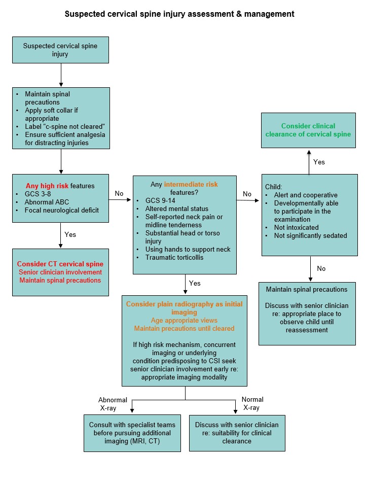

Consider consultation with local specialist teams when
- the cervical spine cannot be cleared clinically
- the role and modality of imaging to investigate suspected CSI is uncertain
- the radiological findings are normal, but the child has persistent symptoms or signs
Note: provision of spinal services varies by region. Utilise appropriate local or tertiary specialist teams as available
Consider transfer when
- all children meeting major trauma criteria, including suspected or confirmed CSI should be referred and transferred to a major trauma service for definitive management. This should be done in consultation with local state pre-hospital and inter-hospital
trauma transfer guidelines
- child requires care beyond the comfort level of the hospital
For emergency advice and paediatric or neonatal ICU transfers, see Retrieval Services
Consider discharge when
- the cervical spine has been cleared
- CSI has been identified and discussed with the appropriate local team. A treatment and follow-up plan is in place
Parent information
Head injury – general advice
Head injury – return to school and sport
Hard collar
NSW Foam Collar Report
Pain relief medicine for Children
Appendix 1: How to apply a soft (foam) collar
- Restrict movement of cervical spine manually
- Provide regular analgesia and reassurance
- Caution should be exercised during moment of risk (Appendix 2)
- Apply foam collar
Measurement and application
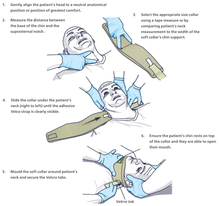
Image used with permission from NSW Agency for Clinical Innovation
Cut along the lower edge of the foam collar to ensure it fits snugly below the chin

 Images used with permission from Children's Health Queensland Hospital and Health Service
Children who are fitted with a pre-hospital collar should be fitted with a new foam collar labelled with “Cervical spine not cleared”

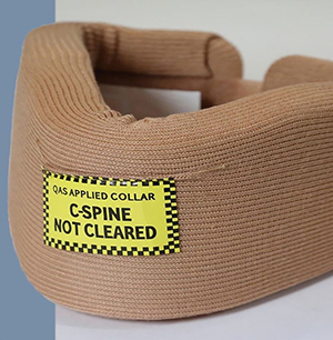
Image used with permission from Queensland Ambulance Service
- If the child is uncooperative or too small to fit a foam collar (baby or infant), movement should be minimised as best as possible without the use of a foam collar. It should be made clear to all staff and family that the cervical spine is not clear
- Note: in children, a collar may cause agitation and potential harm. Distraction of the child and a hands-off technique (eg using a child’s car seat) may be better for maintaining the neck in a position of comfort
Appendix 2: Moments of risk
| Moment of risk |
Strategies to protect cervical spine |
| Trolley transfers and log roll |
Use patslide and have one nurse or doctor dedicated to the head and spine
If
<8 years old, use Thoracic elevation device on arrival |
| Pain/agitation |
Early pain relief
Position caregiver at head of bed to allow direct eye contact and touch
Attendant to reassure and advise family of how to support child
In conscious infants, consider removing collar and allowing child to find
their own best neck position (eg in parent's arms) |
| Vomiting |
Early administration of antiemetics (or gastric tube insertion if appropriate)
Attendant or parent present and "nurse call" for log roll if vomiting |
| Imaging |
Attendant to
- accompany to X-ray if removal of collar or manual re-positioning is required
- ensure TED used appropriately
- ensure that neck neutrality is maintained during CT head and neck
|
| Intubation |
Assign specific attendant to neck positioning
Consider fibreoptic intubation
Avoid unpredictable muscular contractions eg depolarising neuromuscular junction blockade agents |
Appendix 3: Conditions known to predispose to CSI (full list)
- Trisomy 21/Down syndrome (shallower facet joints and hypotonia causing increased risk of horizontal displacement)
- Klippel-Feil syndrome
- Achondroplasia
- Mucopolysaccharidosis
- Ehlers-Danlos syndrome
- Marfan syndrome
- Osteogenesis imperfecta
- Larsen syndrome
- Juvenile rheumatoid arthritis
- Juvenile ankylosing spondylitis
- Renal osteodystrophy
- Rickets
- History of CSI or cervical spine surgery
- Occult congenital deformity such as os odontoideum (a congenitally short odontoid peg limiting the effectiveness of the transverse ligament)
Last updated July 2026