Fracture healing in children follows the same stages as that of adults but occurs at a much faster rate. Fractures heal by forming callus, which follows three overlapping phases: inflammatory, reparative and remodelling.
Phases of fracture healing
Inflammatory phase (duration: hours–days): Broken bones result in torn blood vessels and the formation of a blood clot or haematoma. The inflammatory reaction results in the release of cytokines, growth factors and prostaglandins, all of which are important in healing. The fracture haematoma becomes organised and is then infiltrated by fibrovascular tissue, which forms a matrix for bone formation and primary callus.
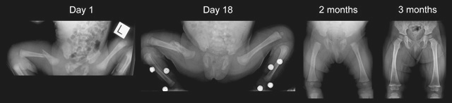
Reparative phase (duration: days-weeks): A thick mass of callus forms around the bone ends, from the fracture haematoma. Bone-forming cells are recruited from several sources to form new bone, which can be seen on radiographs within 7-10 days after injury (Figure 9). Soft callus is organised and remodelled into hard callus over several weeks. Soft callus is plastic and can easily deform or bend if the fracture is not adequately supported. Hard callus is weaker than normal bone but is better able to withstand external forces and equates to the stage of "clinical union", i.e. the fracture is not tender to palpation or with movement.
Remodelling phase (duration: months-years): This is the longest phase and may last for several years. During remodelling, the healed fracture and surrounding callus responds to activity, external forces, functional demands and growth. Bone (external callus) which is no longer needed is removed and the fracture site is smoothed and sculpted until it looks much more normal on an x-ray (Figure 9). The epiphyses gradually realign and residual angulation may be slowly corrected, in accordance with the rules of remodelling, outlined above.

A
B
Figure 9: Young children have excellent remodelling potential. A. These are sequential radiographs of a child who sustained a fracture of the midshaft of the right humerus during birth. Note the very rapid healing with extensive callus by day 7 followed by remodelling. B. These are sequential radiographs of a 14 month child who sustained a fracture of the midshaft of the right femur. Again, note the rapid healing with extensive callus by day 18.