The long bone in a child is divided into four regions: the diaphysis (shaft or primary ossification centre), metaphysis (where the bone flares), physis (or growth plate) and the epiphysis (secondary ossification centre). In the adult, only the metaphysis and diaphysis are present (Figure 1).

Figure 1: Anatomical differences between adult and child bone.
The epiphysis is completely or mostly cartilaginous in infants. Initially it consists of articular cartilage and growth cartilage until these become differentiated by the development of a secondary ossification centre (Figure 2).
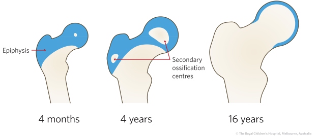
Figure 2: The proximal femoral epiphyseal secondary ossification centre appears at about four months. The ossification centres gradually enlarges until the cartilaginous (blue) area is almost completely replaced by bone at skeletal maturity.
The physis is responsible for the longitudinal growth of long bones whilst circumferential growth is mainly due to periosteal (appositional) growth.
Endosteal reabsorption is part of the growth/remodelling process but it's counterintuitive to say that reabsorption contributes to growth.
Longitudinal and circumferential bone growth
The physis is a barrier to blood flow which may be critical during healing after physeal separations. Understanding which physes, are subject to complete disruption of the blood supply at the time of separation is a guide to the risk of long-term complications such as avascular necrosis (e.g. slipped upper femoral epiphysis).
The physis cannot be seen on x-ray as it is radiolucent but its function can be indirectly appreciated by the assessment of the Harris lines. These fine, radiodense lines occur in the metaphysis of bone and are a normal phenomenon (Figure 3). They are, however, more obvious after illness, injury or administration of certain drugs eg bisphosphonates in children with osteogenesis imperfecta. If the physis is growing normally they should exactly parallel its contour.
Figure 3: Radiographs showing normal Harris lines (arrows) in a child that sustained a Salter-Harris type IV fracture in the distal tibia and Salter-Harris type I fracture in the distal fibula. Note that the Harris line is parallel to the physis, indicating there has been uninterrupted and equal growth of the distal tibia and fibula.