1. Summary
Fractures of the Navicular are Uncommon injuries presenting to an Emergency Department.
A high degree of suspicion may be necessary to establish the diagnosis.
Children presenting with exacerbation of medial foot pain need to have other differential diagnoses considered, including Köhler disease and Accessory Navicular
2. How are they classified?
- By part of the navicular affected (body, tuberosity or avulsion).
- Acute traumatic fractures or stress fractures.
3. How common are they and how do they occur?
Navicular Fracture is an infrequent presentation in paediatric practice.
Usual mechanism:
- navicular body fractures
- axial loading
- Usually high-impact mechanism such as motor vehicle accident or high fall
- navicular tuberosity fractures
- eversion with simultaneous contraction of Posterior Tibial Tendon (often associated with accessory navicular)
- navicular avulsion fractures a.k.a dorsal avulsion or tuberosity avulsion
- plantarflexion or eversion/inversion
- often a low-energy injury
- stress fracture
- chronic overuse / running on hard surfaces.
4. What do they look like – clinically?
Patients with acute fractures of the navicular usually present with:
- Pain over the dorsal or dorsomedial foot following trauma.
- Localized swelling and tenderness over the navicular.
- Difficulty with weight-bearing
- Increased pain and inability to walk on or push off (ie, plantarflex while weight-bearing) with their toes.
The differential diagnosis of Köhler Disease (Avascular Necrosis of the Navicular) is suggested more by chronic progressive medial midfoot pain worse with weight-bearing, though it can sometimes present with acute onset of symptoms. There is usually swelling and tenderness over the navicular, with the patient preferring to bear weight through the lateral part of the foot. X-Ray appearance and management are given below. |
Chronic or progressive medial midfoot pain may also be caused by Accessory Navicular. This is a developmental anomaly in which the tuberosity of the navicular develops as a secondary ossification centre but fails to unite with the main navicular.
Whilst asymptomatic in many, pressure from inappropriately narrow footwear can cause chronic pain and tenderness in the medial midfoot with swelling and erythema, with symptoms usually worse with weight-bearing. X-Ray appearance and management are given below. In symptomatic accessory navicular, resisted strength testing of posterior tibial tendon often elicits pain (have the patient invert and plantar flex the foot against resistance)
|
5. What radiological investigations should be ordered?
AP, lateral and oblique views of the foot
CT may be indicated where a high degree of suspicion persists in the face of apparently normal radiographs. This warrants orthopaedic consultation, either on the day of presentation or in the fracture clinic after a period of immobilisation.
6. What do they look like on x-ray?
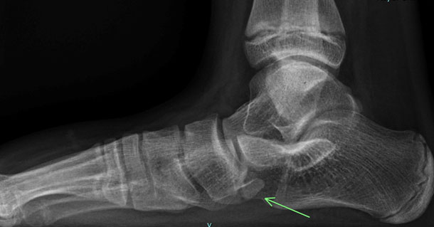
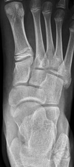
 Dorsal avulsion fracture
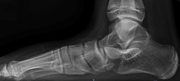
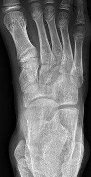
 Navicular Body fracture
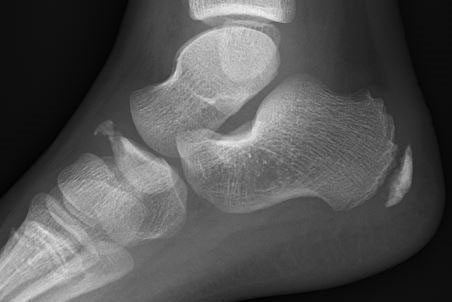
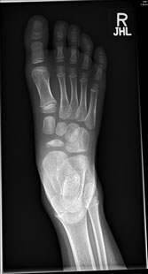 Kohler disease, with flattening and sclerosis of the Navicular Kohler disease, with flattening and sclerosis of the Navicular
|
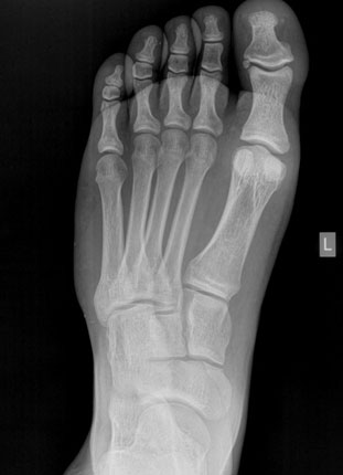
Accessory Navicular shown on the first image, with normal image shown at right for comparison.
|
7. When is reduction (non-operative and operative) required?
Not indicated in the ED.
8. Do I need to refer to orthopaedics now?
Immediate orthopaedic referral required where there is
- displacement of the fracture
- a suspected or confirmed LisFranc injury
- open fracture.
Other Navicular fractures can simply be reviewed in the fracture clinic
9. What is the usual ED management for this injury, and what follow-up is required?
- Immobilise fractures in a below-knee splint or cast
- Advise the child and parent to avoid bearing weight until orthopaedic review in approximately one week
Köhler Syndrome usually has below-knee immobilisation (eg a CAM-boot) applied for 4-6 weeks with follow-up arranged with a sports medicine physician. Long-term outcomes of non-operative management are almost universally good in children - surgical treatment is very rarely indicated, though symptoms can take some months to resolve. Ongoing symptoms after 6 weeks of immobilisation is an indication for orthopaedic referral. |
First-line management of symptomatic accessory navicular is nonoperative.
For significant symptoms, immobilise in a CAM boot for 6 weeks; in mild symptoms aim to relieve pressure by avoiding narrow footwear and/or footwear modification, and consider a period of short-term activity modification. Orthopaedic follow-up is for those who have ongoing symptoms despite nonoperative management; surgical management is occasionally indicated in older children or adolescents. |
10. What advice should I give to parents?
Keep the limb elevated where possible in the first few days after injury to prevent swelling
11. What are the potential complications associated with this injury?
Complications are uncommon in non-displaced fractures.
12. References
Chan J & Young J, Köhler Disease: Avascular Necrosis in the Child, Foot Ankle Clin, 2019;24(1):83-88
Wynn, M et al, Effectiveness of nonoperative treatment of the symptomatic accessory navicular in pediatric patients, Iowa Orthopaedic Journal 2019;39(1),45-49
Leonard, Z et al, Adolescent Accessory Navicular, Foot Ankle Clin, 2010;15(2):337-347